I wanted to explain how structural and functional MRI image format NIFTI works. NIFTI stands for Neuroimaging Informatics Technology Initiative. It is currently a 8 to 128 complete bit, signed or unsigned integer data storage format. The most common implementation of NIFTI is the 16 bit signed integer data storage. The NIFTI images usually have the file extensions *.nii or (*.hdr and *.img) pairs. This image format is important due to two reasons: 1) The format stores the spatio-temporal imaging details and 2) compression to allow better space management.
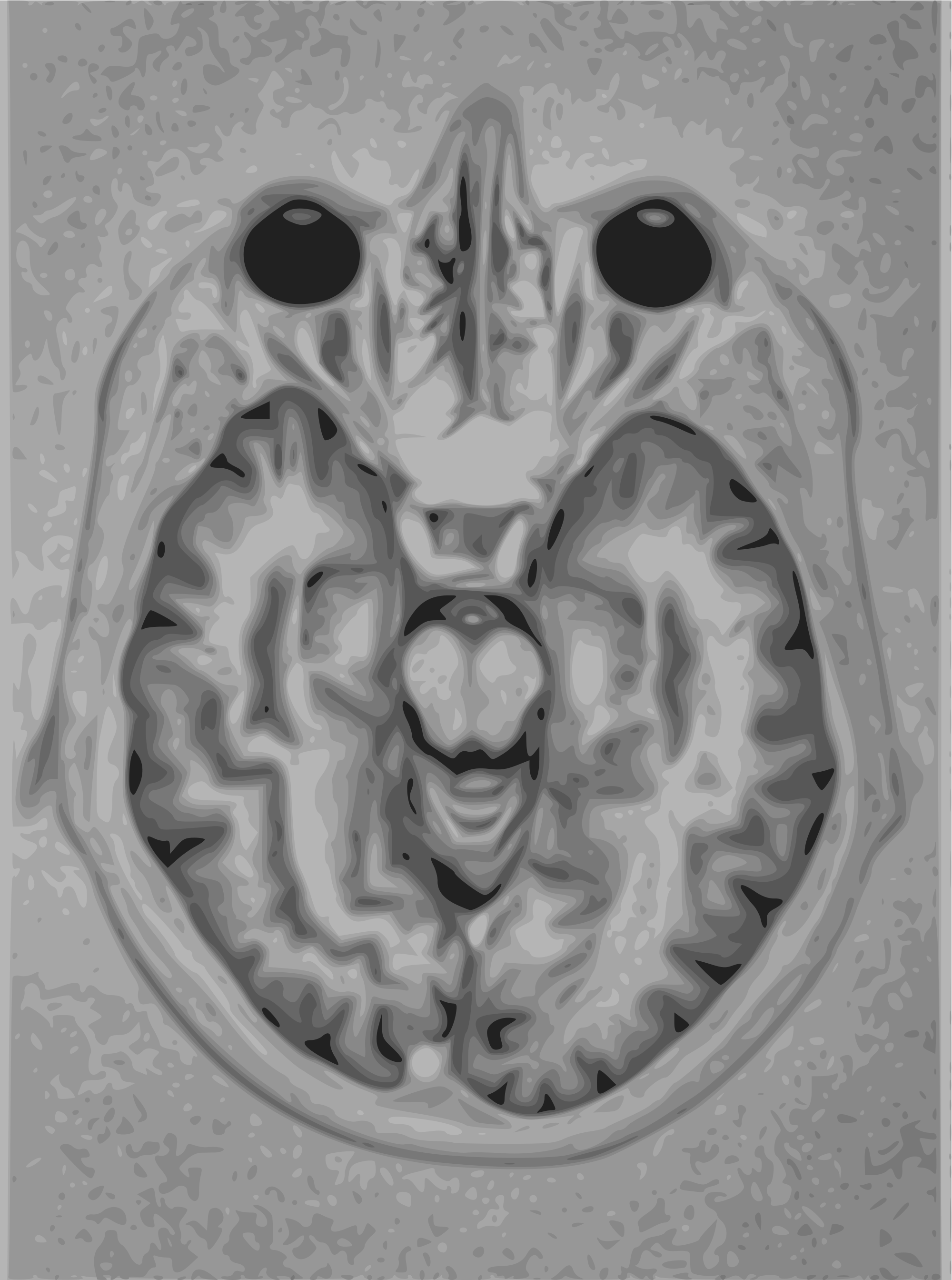
Anyone who underwent a brain scan knows, the picture of the brain from an MRI or CT scanner is usually a grayscale picture with 65,536 shades of gray. The raw files from the scanner are usually DICOM (Digital Imaging and Communications in Medicine) format with an extension of *.dcm. The DICOM format is similar to the RAW image format for cameras. Instead of pixel read out stored in RAW images, the DICOM images store scanner readouts.
Each scan of a subject usually contains several DICOM files. This is an advantage and a disadvantage. For sharing specific image slices, DICOM is therefore extremely useful. But, for most interpretation purposes, the analysis often requires full image sets. A few slices from the scanner becomes less useful. This is where NIFTI format comes to rescue.
Since the format stores the entire sequence in a single file, the issues of managing large number of files are eliminated. The interpretation of a specific image based on the image preceding and succeeding it becomes easier. This is due to the ordered arrangement of images.
There is another important advantage of NIFTI. Brain imaging data is most relevant from an analytical point of view, to be used as a 3D data structure. Even though the individual components of the NIFTI are 2D images, the interpretation of an image becomes more reproducible if we treat them as 3D images. For this purpose, the NIFTI format is the best format to work with.
An example is the use of a machine learning tool called 3D convolutional neural networks (cnn). 3dcnn’s provide the 3d spatial context to a voxel. For image sequences like brain scans, identification of various structures or any abnormalities require the 3d spatial context of a voxel. The 3d cnn approach is very similar to looking at a video and trying to identify what the scene is about. Instead of using it for video scene recognition, 3d cnn can be used to train and detect specific features in a brain scan.
This work is done as part of our startup project nanoveda. For continuing nanoveda’s wonderful work, we are running a crowdfunding campaign using gofundme’s awesome platform. Donation or not, please share our crowdfunding campaign and support our cause.
Donate here: gofundme page for nanoveda.